CBCT Question Of The Month
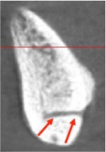
In the midline of the mandible, this radiolucent canal (red arrows) is often seen on cross-sectional images. What is this radiolucent canal called? What possible complication may arise from placing an implant in this area?
Prostho Question Of The Month
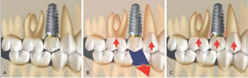
When inserting a dental implant crown with adjacent natural teeth, ideally, a “timed occlusal” scheme should be utilized. What exactly is “timed occlusion”?
Implant Study Of The Month
In evaluating the biofilm (plaque) that accumulates on various implant materials, a study compared two common implant restorative materials (porcelain and zirconia) on the;
- Percent of the material that coated with biofilm
- The thickness of the biofilm

Which material was shown to be superior in preventing biofilm accumulation?
Answers Below –
Answers
1.) The radiolucent canal in the mandibular midline is termed the lingual vascular canal (LVC) or mandibular vascular canal and contains the right and left sublingual arteries. In theory, implant osteotomy preparation in this area may lead to significant intraosseous bleeding episodes which may be controlled by placing a direction indicator, surgical drill, or the implant into the osteotomy site to allow for the clotting process.
2.)
A.) In light occlusion, no contact should exist on the implant prosthesis as thin articulating paper or shimstock (approximately 10 μ m) is easily pulled through.
B.) On heavy occlusion(clenching), the teeth will move apically (periodontal ligament), and the implant crown will have light contact (i.e., shimstock having resistance when pulled through). C.) If implant prosthesis and natural teeth occlude evenly with light occlusion, the implant will be in hyperocclusion and subject to biomechanical overload.
3.) The lowest surface coating (19.0%) and biofilm thickness (1.9 μm) were determined with zirconia, while the highest mean values were identified with porcelain ceramics (46.8%, 12.6 μm)
Bremer, Felicia, et al. “In vivo biofilm formation on different dental ceramics.” Quintessence International 42.7 (2011).